
Mellon Grant African Art Conservation Process
A look at the process behind art conservation and the tools we use to help us.
Tools
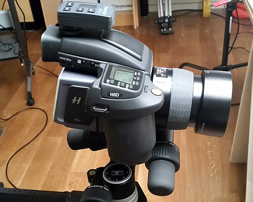 Our H6D Hasselblad DSLR
 Imaging Equipment
Technique: Digital imaging/digital photography
Equipment: Hasselblad H6D Medium Format DSLR Camera
What it tells us: Digital imaging is the most valuable tool we have for visually documenting objects. With its 100MP Sensor, the Hasselblad produces images in such high resolution that we can zoom in on details only otherwise visible under the stereomicroscope.
What it will help us analyze: All objects in this study will be documented through Digital imaging, allowing us to study the objects in further detail and share information with scientists and other colleagues without having to move or manipulate the objects themselves.
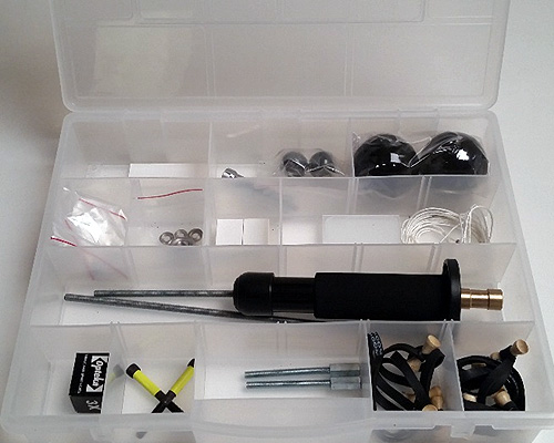 Our RTI Capture kit
 Technique: Reflectance Transformation Imaging (RTI)
Equipment: RTI Capture Kit from CHI
What it tells us: RTI is a computational imaging technique that provides us with detailed visual information about surface morphology and texture. Through this method we can capture an object’s surface shape and color, creating a permanent record that can be manipulated from the RTI software to allow interactive re-lighting from any direction, helping to highlight features of interest.
What it will help us analyze: RTI will be useful for looking at objects with hard to see surface texture, patterns, or lettering.
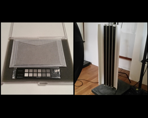 (left) UV Innovations Ultraviolet Photography Standard, (right) UV Studio lights
 Technique: Ultra Violet induced Fluorescence (UV)
Equipment: Range of long and short wave UV lights coupled with our DSLR camera (listed above)
What it tells us: The characteristic fluorescence of different materials helps us observe otherwise invisible coatings and materials on the surfaces of objects. In some cases the characteristic color of fluorescence may help us identify the material.
What it will help us analyze: Objects with past restorations, coatings, and fluorescent materials not discernible under normal lighting conditions.
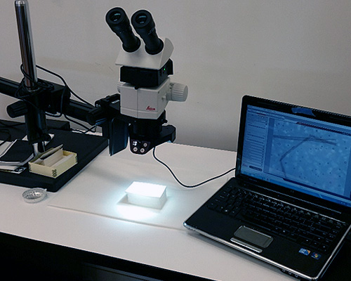 Stereomicroscopy setup
 Technique: Stereo microscopy
Equipment: Leica M80 Microscope with Leica IC80HD Camera
What it tells us: High magnification imaging helps us understand the structure of materials on a microscopic scale and document these features. We can also share the captured images with scientists and other colleagues to help identify materials found on our collection objects.
What it will help us analyze: Everything that can fit under the microscope!
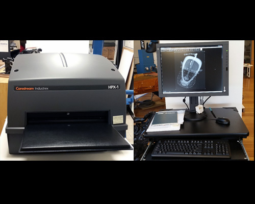 (Left) Carestream scanner and (right) X-radiography computer workstation
 X-Radiography Equipment
Technique: Digital (computed) X-radiography
Equipment: Carestream HPX-1 digital scanner and phosphor plates
What it tells us: X-radiography allows us to produce an image of the internal structure of an object, and in many cases allows visualization and identification of hidden materials and construction methods. Computed radiography allows us to employ a variety of processing techniques instantaneously through the software to help reveal additional details and features of interest. These files can also be shared with colleagues easily as digital image files.
What it will help us analyze: Objects with parts joined together, an internal frame or structure, or objects that cannot be opened and which contain unknown materials (e.g. a wooden sculpture with an enclosed chamber).
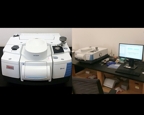 The Thermo Nicolet IS50 with Raman module inserted and FTIR computer workstation
 Technique: FT-IR and Raman
Equipment: Thermo Nicolet IS50 FTIR with Raman module
What it tells us: FTIR helps distinguish the molecular structure of materials due to their characteristic absorption of IR light. A spectral “fingerprint” is produced when a sample is run and can then be compared to known reference samples in order to help identify the unknown. See here for more details. Raman spectroscopy similarly produces a molecular fingerprint based on the interaction of a laser beam with the sample material.
What it will help us analyze: FTIR and Raman will be useful in identifying organic materials such as resins, adhesives, and plastics, as well as inorganic materials including some pigments and minerals.
